Glaucoma

What is Glaucoma?
Glaucoma is a disease that results in damage to your eye’s optic nerve: which is the nerve leading to the brain that controls your vision. Glaucoma usually develops when fluid builds up in the front part of your eye causing the pressure in your eye to increase above normal levels. This elevated pressure can injure your optic nerve, and cause vision loss.
There Are Two Major Types of Glaucoma
Primary Open-Angle Glaucoma
Primary Open-Angle Glaucoma (POAG) is the most common type of glaucoma. This type of glaucoma develops gradually over time when the eye doesn’t drain fluid as well as it should.
Similar to water backing up in a sink with a clogged drain, the pressure in the eye can slowly build up and cause damage to the optic nerve. This type of glaucoma causes few symptoms, is often painless, and early on causes few vision changes. It is often referred to as the “sneak thief of sight.” By the time vision loss is noticed by the patient, there can be significant, irreversible damage to the optic nerve.
Normal Tension Glaucoma is another type of Open-Angle Glaucoma in which the eye pressure stays within the normal range, but still results in damage to the optic nerve.
The treatment for this type of glaucoma is similar to Primary Open-Angle Glaucoma. But if often requires a medical evaluation to make sure that no other conditions are causing damage to the nerve and the vision.
Angle-Closure Glaucoma
Angle-Closure Glaucoma occurs when the iris (the colored part of your eye) sits too close to the drainage angle in your eye. The iris ends up mechanically blocking the drainage angle, so that fluid cannot escape from the eye. If the iris completely blocks the angle, it can cause an Acute Angle-Closure attack, where the eye pressure increases quickly and can cause serious damage to the optic nerve in a short period of time.
Symptoms of Acute Angle-Closure Glaucoma include:
- Headache
- Sudden blurry vision
- Severe eye pain
- Nausea and vomiting
- Halos around lights
If you have any of these symptoms you should call your Ophthalmologist immediately to receive treatment to avoid losing vision or going blind in your eye.
How is Glaucoma diagnosed?
During a comprehensive eye exam, Dr. Gamell will evaluate the overall health of your eye, such as measuring the pressure in your eye and assessing the drainage angle with a test called gonioscopy. She will also examine your optic nerve to identify existing damage. Photographs of your optic nerve, along with special diagnostic testing can help determine the presence of and/or stage of disease. Typically, Dr. Gamell will order a Humphrey Visual Field, which can detect a loss of peripheral or side vison. In addition, she will obtain Optical Coherence Tomography or OCT which can detect very early damage to the optic nerve. Dr. Gamell uses these tests results, along with findings during your eye exam to help determine if you have glaucoma.
Based on the results of your exam, Dr. Gamell takes time to discuss your options for treatment and works closely with you to make the right decision regarding care of your eyes.
How is Glaucoma treated?
Once you develop glaucoma damage, it is permanent and cannot be reversed. Therefore, early diagnosis and treatment is important. There are many different ways to treat glaucoma. Typically, eyedrops or laser are used as initial therapy to control eye pressure. However, a combination of medical, laser and incisional surgery may be needed at various points during your lifetime to control the disease. Glaucoma in that way is similar to Diabetes; it is a chronic disease that might not be cured, but can usually be controlled to help preserve your vision.
Eyedrop Medications
Medicated eye drops are available for lowering pressure in your eye by reducing the amount of fluid it produces. There are many different types of eyedrops available now. Depending upon your medical history, Dr. Gamell will help you choose the best medicine or combination of medicines for you.
Laser Surgery
There are several types of laser surgery available to help treat glaucoma. The type of laser therapy Dr. Gamell recommends depends upon the type and severity of the glaucoma you have.
Laser Iridotomy

What is a Laser Iridotomy?
A laser iridotomy is the standard treatment for Narrow-Angle or Closed-Angle Glaucoma. This laser procedure can also prevent further damage to the optic nerve that results from the condition.
The goal a Laser Iridotomy is to increase the efficiency of fluid drainage from the eye to help you maintain normal pressure.
Why do I need an iridotomy?
In Narrow Angle Glaucoma, the drainage angle is blocked or closed in certain areas. The angle is the space between your cornea (the clear part of your eye) and the iris (the colored part) that contains the trabecular meshwork that drains fluid from the eye.
When something blocks the meshwork, it causes fluid to build up in your eye and put pressure on your optic nerve. Without treatment, this type of glaucoma can lead to irreversible blindness.
If you have a narrow angle, Dr. Gamell may recommend a laser iridotomy to help prevent damage to your optic nerve. In addition, if the eye pressure is elevated and there is evidence of glaucomatous damage, she may recommend an iridotomy to help prevent further glaucoma damage. During your exam, Dr. Gamell will discuss all the best options for managing your disease.
What happens during a Laser Iridotomy?
A Laser Iridotomy is performed either in the Ophthalmologist office or an Ambulatory Surgery Center. You are given eye drops to make your pupil small. Then a topical eyedrop is used for anesthesia to numb your eye. A contact lens in placed on your eye and a laser is used to make a small hole in the edge of your iris. This hold is not visible to the naked eye and can usually only be seen when your doctor examines you with a slit lamp microscope in the office. The procedure itself takes only a few minutes, but the office visit may take an 1 ½ to 2 hours, since you have to wait to have your pressure rechecked an hour after the procedure.
After the laser procedure, you will be prescribed eyedrops for a few days. Dr. Gamell rechecks your eye a few weeks later.
Argon Laser Peripheral Iridoplasty
In patients who have Narrow Angles, a laser iridotomy is usually the first procedure performed to help open up the angle and prevent an angle closure attack. In some patients, however, the angle does not adequately open after a Laser Iridotomy. In certain cases, Argon Laser Peripheral Iridoplasty may be performed to help open the angle further.
What happens during Argon Laser Peripheral Iridoplasty
Argon Laser Peripheral Iridoplasty is performed either in the Ophthalmologist office. You are given eye drops to make your pupil small. Then a topical eyedrop is used for anesthesia to numb your eye. A contact lens in placed on your eye and a laser is used applied to the edge of your iris to help pull the iris away from the angle. The procedure itself takes only a few minutes, but the office visit may take an 1 ½ to 2 hours, since you have to wait to have your pressure rechecked an hour after the procedure.
After the laser procedure, you will be prescribed eyedrops for about a week. Dr. Gamell rechecks your eye a few weeks later. If the Argon Laser Peripheral Iridoplasty does not improve the appearance of your angle, Dr. Gamell may suggest cataract surgery in the future.
Selective Laser Trabeculoplasty (SLT)
What is SLT?
Selective Laser Trabecuoplasty or SLT is a type of laser procedure that treats Open Angle Glaucoma.
In Open Angle Glaucoma, age related changes can cause the natural drainage system of your eye (the trabecular meshwork) to not function well. Fluid doesn’t drain properly from the eye and causes a rise in eye pressure. During SLT, the trabecular meshwork is treated with a laser to help the eye drain fluid more effectively and lower the eye pressure.
Why do I need SLT?
If you have Primary Open Angle Glaucoma or Secondary Open Angle Glaucoma, you may be a candidate for SLT. SLT may be used in combination with eyedrops to help lower your eye pressure to the desired level, or your “goal eye pressure.” If you have multiple allergies to eyedrops, SLT may help decrease your dependence upon eyedrops to lower your eye pressure. In addition, SLT is approved as first line therapy for glaucoma, and can even be used initially in place of eyedrops. If you are interested in SLT, schedule a consultation with Dr. Gamell and she can help you explore your options.
How effective is SLT?
After SLT, the eye pressure can drop anywhere from 20-30%. The results can last from 2-5 years. In addition, the procedure can be repeated even if the initial result has worn off. If you have already had SLT and are wondering if you would benefit from additional treatment, you can schedule a consultation with Dr. Gamell.
There are also other laser procedures available that may help control your eye pressure.
Micropulse Laser Cyclophotocoagulation
What is Micropulse Laser Cyclophotocoagulation (Micropulse CPC)?
Micropulse Laser Cyclophotocoagulation (Micropulse CPC) is a laser procedure that is usually performed in an Ambulatory Surgery Center. A special probe is applied to the outside of your eye for 1-2 minutes. Usually, mild sedation is given to help with pain control for Micropulse CPC.
You will take special eyedrops after the procedure to control inflammation. After about 6-10 weeks, the final effect of this laser procedure can be seen. Often patients can see good pressure control and even stop using some of their eye drop medications after this procedure.
Diode Transcleral Cyclophotocoagulation
Diode Transcleral Cyclophotocoagulation (or Diode TSCPC) is performed with the same laser unit as the Micropulse TS CPC procedure. However, a different probe is used, as well as different laser settings that make this procedure better suited for more advanced stages of disease. Like Micropulse TS CPC, the Diode TS CPC is a laser procedure that is usually performed in an Ambulatory Surgery Center. You are usually given mild sedation along with a local injection for pain control during the procedure. A special probe is applied to the outside of your eye for a few minutes. Then you are sent home with an eye patch on your eye.
You will take special eyedrops after the procedure along with your glaucoma eyedrop medications. After about 6-10 weeks, the final effect of this laser procedure can be seen. Often patients can see good pressure control and even stop using some of their eye drop medications after this procedure.
Minimally Invasive Glaucoma Surgery (MIGS)
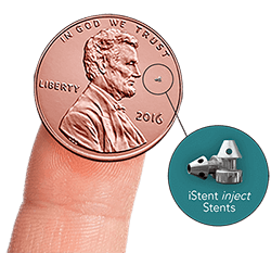
Minimally Invasive Glaucoma Surgeries describe a group of surgeries that are less invasive and shorter in duration than traditional procedures such as Trabeculectomy and Tube Shunt procedures. In many cases they also carry with them a lower risk of complications.
These procedures can either be performed at the time of Cataract Surgery, or in certain cases on their own without Cataract Surgery.
If you are interested in MIGS surgery as an option, make an appointment with Dr. Gamell for a glaucoma evaluation. She can discuss your various options with you.
Trabeculectomy
A trabeculectomy is a traditional surgical technique to create a flap in the outer layer of the eye to promote fluid drainage. This surgery involves creating a pocket known as a filtration bleb that allows surrounding tissue to absorb extra fluids to lower eye pressure. Trabeculectomy surgery can be very effective in stabilizing eye pressure. Dr. Gamell can help you determine if this type of surgery is right for you.
Glaucoma Drainage Implants
A glaucoma drainage implant is another option to help control eye pressure. This procedure can be useful in many different types of glaucoma; but is particularly helpful in patients with a history of trauma, eye inflammation, neovascular glaucoma due to diabetes or a stroke in the eye, and other eye disorders. There are different types of glaucoma drainage devices available. In these procedures, a tube is inserted into the angle of the eye which drains fluid and controls the eye pressure. The fluid collects under the conjunctiva of the eye near the base or plate of the implant device. These implants can be very effective in lowering pressure, especially in cases where a trabeculectomy or laser surgery is not appropriate. Dr. Gamell can help you decide which type of Glaucoma Drainage Device is right for you.
Glaucoma Surgery Follow Up Care
For glaucoma incisional surgeries, the post-operative care can almost be as important as the surgery itself. Post-operative visits help ensure that your eye heals properly, the eye pressure remains controlled, and that you do not develop an eye infection. You will be on varying doses of post-op eyedrops medications, and decisions about your glaucoma medications will be made at the post-operative visits.
For most surgeries, patients will be seen at least 1 day, 1 week and 1 month after their procedure. Depending upon the type of surgery, more post-operative visits may be required. Ensuring that you can follow-up for your post-operative exams is critical, so be sure to coordinate with Dr. Gamell when you schedule your surgery. If you have plans to travel, we recommend you wait at least a month after your MIGS or Incisional Glaucoma Surgery procedure.